
Scanning Probe Microscopy
Scanning probe microscopes for investigating surface topography, nanostructures, and local physical properties of materials.
What Is Scanning Probe Microscopy?
Scanning Probe Microscopy (SPM) is a family of techniques used to investigate material surfaces with a highly sensitive probe that scans a selected area of a specimen. Unlike optical and electron microscopy, surface images are not formed directly using light or an electron beam. Instead, they are generated by measuring physical interactions between the probe tip and the material under investigation.
Scanning probe microscopes are used to study surface topography, roughness, microstructures and nanostructures, as well as local mechanical, electrical, and magnetic properties when suitable measurement modes are available. Applications include materials science, nanotechnology, microelectronics, polymers, thin films, coatings, biophysics, and research laboratories.
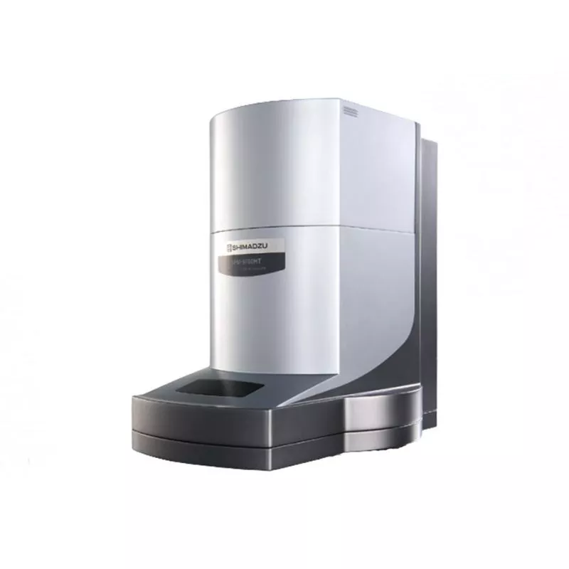
One of the most widely used SPM techniques is Atomic Force Microscopy (AFM). It allows researchers to investigate conducting and nonconducting surfaces and acquire three-dimensional topographic data. This category includes Shimadzu scanning probe microscope systems, including the SPM-9700HT for high-resolution surface characterization.
Operating Principle of Scanning Probe Microscopes
The principal sensing element of an atomic force microscope is a microscopic cantilever carrying a sharp probe tip. During scanning, interactions between the tip and specimen cause changes in cantilever deflection or oscillation behavior. These changes are detected and processed to generate information about the surface.
Many AFM instruments use an optical lever system to measure cantilever deflection. A laser beam is reflected from the cantilever onto a photodetector. A precision positioning system moves the probe or specimen through a selected scan area, while software generates height maps and other measured parameter distributions.
Depending on the interaction with the specimen, scanning probe microscopes may operate in contact, dynamic, or other specialized modes. The result is a digital image or map representing a particular surface characteristic.
Spatial resolution depends not only on the measurement system but also on tip geometry, specimen condition, scan parameters, and operating mode. Quantitative measurements of roughness, feature height, and local properties require appropriate calibration and data processing.
Atomic Force Microscopy (AFM) and Measurement Modes
Atomic Force Microscopy characterizes surface topography and properties by measuring interactions between a cantilever tip and the specimen. Different scanning modes provide complementary information about surface structure and local behavior.
- Contact Mode — the probe tip remains in controlled mechanical contact with the surface. This mode is used for topographic imaging and analysis of suitable rigid specimens.
- Dynamic Mode — the cantilever oscillates near its resonance frequency, and changes in oscillation behavior are detected as the probe approaches the surface. Under suitable conditions, this can reduce mechanical interaction compared with contact scanning.
- Phase Mode — measures changes in the phase of cantilever oscillation, revealing contrast associated with differences in local material properties, including heterogeneity in polymers and composites.
- Lateral Force Microscopy (LFM) — measures cantilever torsion associated with lateral forces during surface contact and provides information about friction-related contrast.
- Force Modulation Mode — applies periodic mechanical excitation to investigate variations in local deformation response.
- Force Curve Measurements — record probe-sample interactions as a function of separation distance and may be used to investigate adhesion and other local mechanical characteristics with suitable procedures.
Beyond standard AFM modes, specialized systems may support electrical current mapping, surface potential measurements, magnetic force imaging, and other techniques. Availability depends on instrument configuration, probe type, accessories, and software.
Surface Topography, Roughness, and Local Property Analysis
A major application of scanning probe microscopy is the characterization of microscopic and nanoscopic surface relief. Scanning data can reveal protrusions, depressions, particles, grains, structural heterogeneity, defects, and changes caused by manufacturing processes.
Digital height maps can be processed to determine surface profiles, local height differences, roughness parameters, and other geometrical properties. Meaningful comparison requires consideration of scan size, resolution, filtering, image leveling, and probe tip geometry.
Specialized operating modes extend the analysis beyond surface shape. Depending on the configuration, they can generate distributions of lateral forces, phase contrast, surface potential, electrical current, and magnetic interactions. Such measurements are useful for heterogeneous materials, thin films, multiphase polymers, functional coatings, and microelectronic structures.
Scanning probe microscopy is not a direct technique for determining elemental or molecular composition. X-ray fluorescence, electron probe microanalysis, spectroscopy, and other analytical methods may be required for chemical characterization.
Shimadzu SPM-9700HT Scanning Probe Microscope
Shimadzu SPM-9700HT is a high-resolution scanning probe microscope designed for investigating surface topography and local physical characteristics. It uses atomic force measurement techniques and supports digital acquisition and analysis of three-dimensional surface images.
The standard configuration supports Contact, Dynamic, Phase, Lateral Force, and Force Modulation modes. Additional options may provide electrical current mapping, surface potential analysis, magnetic force imaging, and specialized local property mapping.
The SPM-9700HT incorporates a sensitive cantilever displacement detection system and a scanner optimized for efficient image acquisition. Its software supports surface visualization, cross-sectional profiling, three-dimensional rendering, and processing of measurement data.
The instrument is used to investigate metallic and polymer surfaces, functional coatings, thin films, nanostructures, and other materials. Available operating modes, scan ranges, and analysis features depend on the installed scanner and instrument configuration.
Applications of Scanning Probe Microscopy
SPM and AFM methods are used in industrial and research laboratories requiring high spatial detail in surface topography and local property measurements.
- Materials Science — study of microstructures and nanostructures, grains, defects, phase heterogeneity, and surface morphology.
- Nanotechnology — investigation of nanoparticles, nanostructured coatings, thin layers, and functional material surfaces.
- Electronics and Microelectronics — characterization of semiconductor structures, substrates, thin films, and local electrical properties with suitable measurement modes.
- Polymer Manufacturing — examination of polymer morphology, composites, phase heterogeneity, coatings, and surface changes following processing.
- Metallurgy and Mechanical Engineering — investigation of metal surface relief, coatings, machining marks, and roughness characteristics.
- Biomaterials and Biophysics — research on suitable biological and biocompatible surfaces, structural features, and local mechanical interactions.
- Research Laboratories — new material characterization, comparative surface studies, development of nanoscale measurement procedures, and evaluation of manufacturing processes.
How to Choose a Scanning Probe Microscope
Selecting scanning probe microscopy equipment begins with specimen type, scanning area, required spatial resolution, surface characteristics, and the properties to be measured.
Key criteria include supported measurement modes, available cantilevers and probe tips, scanner travel ranges, positioning stability, image acquisition speed, analysis software, and compatibility with optional modules.
For soft polymers and biological materials, probe-sample interaction forces require particular attention. Electrical or magnetic measurements need suitable probes and operating modes. High-resolution measurements may also require vibration isolation, thermal stability, and carefully prepared specimen surfaces.
A properly selected scanning probe microscope provides reproducible information about surface topography, nanostructures, and local material properties and complements optical microscopy, electron microscopy, and spectroscopic analysis.